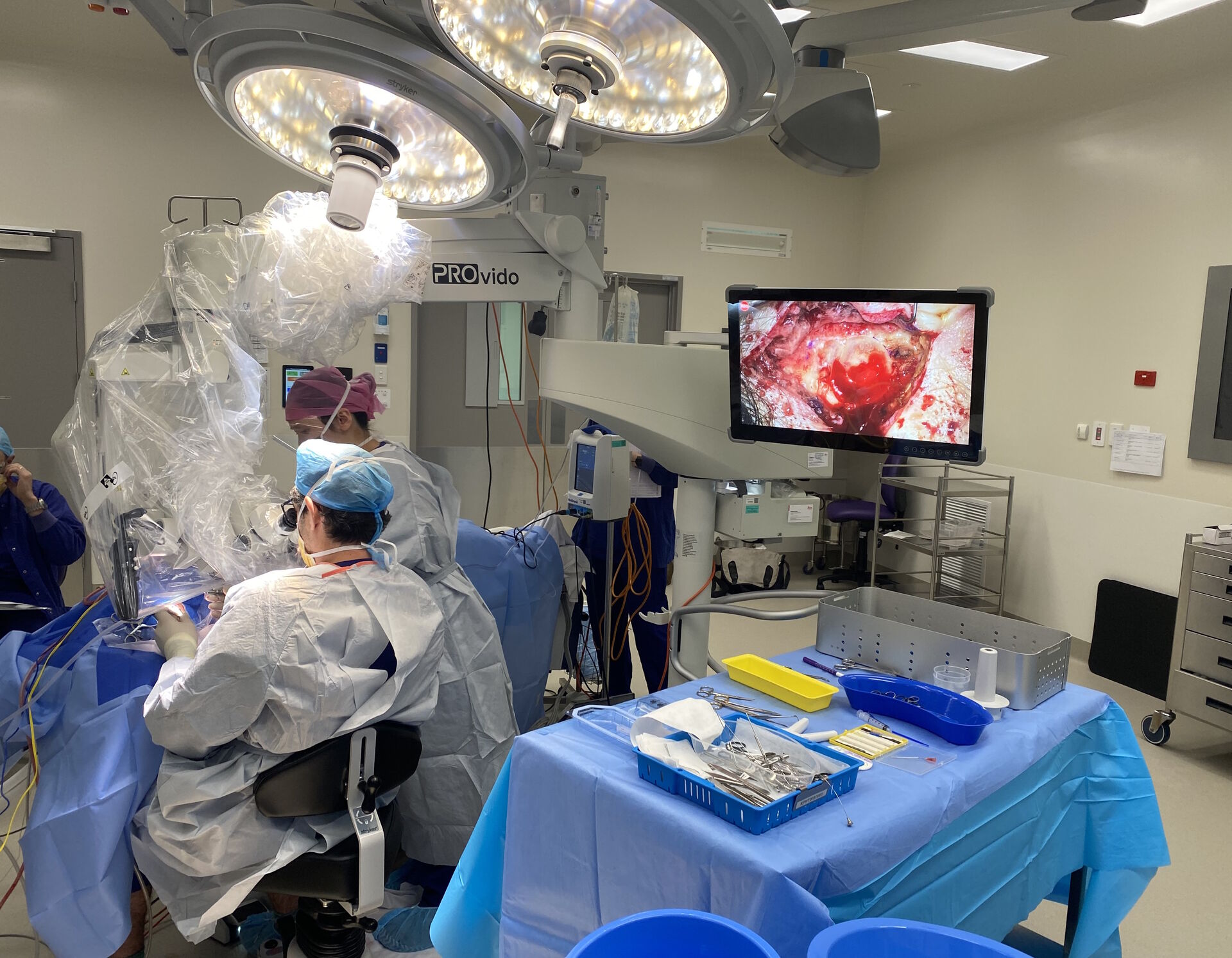

In the following tympanoplasty surgery cases, Dr. Sean Flanagan demonstrates the three standard approaches: post-auricular, endaural and transcanal. He shows how the PROvido microscope, with its innovative technologies, offers a deep, bright view in full focus and allows bimanual manipulation of the tissues. He also highlights its versatility and manoeuvrability supporting smooth surgery.
Case 1: Myringoplasty with a post-auricular approach
The first tympanoplasty surgery case is an anteriorly based perforation of the left ear, with surrounding myringosclerosis. Myringosclerosis is a type of tympanosclerosis involving only the tympanic membrane. Due to the anterior aspect of the perforation, a post-auricular approach is chosen.
The limited post-auricular incision offers excellent cosmesis, but also maximum visualization. After graft harvesting, a triangular periosteal incision is performed, into the zygomatic group, allowing the use of a Freer dissector to elevate the soft tissue, in and around the external auditory canal.
The placement of a Fisch articulated self-retainer allows tension to facilitate dissection. After the hemitransection of the canal and rim excision with a Rosen needle, the tympanomeatal flap is raised using a round knife to identify the tympanic annulus.

The incision of the middle ear mucosa with a Rosen needle allows the elevation of the annulus. The surgeon identifies the chorda tympani nerve, before inserting the temporalis fascia graft and replacing the tympanic membrane.


Case 2: Myringoplasty with an endaural approach
The second tympanoplasty surgery case illustrates a posteriorly based left perforation, with the use of an endaural approach. The pre-operative photograph shows an irregular margin at the anterior aspect and myringosclerosis on the posterior margin.
An initial limited endaural incision is performed, with diathermy dissection down to bone. A tragal incision allows the harvesting of compound perichondrial and cartilage graft. To support this step, the PROvido microscope provides high resolution visualization and an enhanced depth of field.
Two double pronged curved self-retainers are placed perpendicular to each other, before raising the tympanomeatal flap. After sizing the perforation, the surgeon places the composite perichondrial and cartilage island graft. He then places a silastic sheet and gelfoam with antibiotic ointment.


The final step of the tympanoplasty procedure is the closure of the temporalis muscle and skin.
Case 3: Myringoplasty with a transcanal approach
The third tympanoplasty surgery case further illustrates the versatility of the PROvido surgical microscope, with a transcanal approach used to perform a tuck through graft. Even through a speculum, excellent visualization is achieved, according to Dr. Flanagan.
The edges of the perforation are freshened with a Rosen needle. Gelfoam is used for packing to the middle ear prior to placing a perichondrial graft.
Adjustments are made to the graft to ensure appropriate underlay. Silastic sheeting is also used to create surface tension and allow appropriate packing of the canal with gelfoam and Kenacomb ointment.
To discover the full tympanoplasty surgery cases, watch the video:
Disclaimer: Please note that off-label uses of products may be discussed. Consult with regulatory affairs for cleared indications for use in your region. The statements of the healthcare professionals included in this presentation reflect only their opinion and personal experience. They do not necessarily reflect the opinion of any institution with whom they are affiliated or Leica Microsystems.
 performing ear, nose and throat (ENT) surgery using the MyVeo surgical visualization headset.")

 or Minor’s syndrome.")
